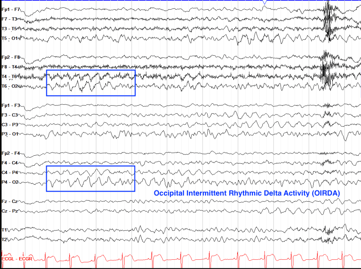
EEG Sample: Learning EEG
Yamada, Thoru, and Elizabeth Meng. Practical Guide for Clinical Neurophysiologic Testing: EEG. Available from: Wolters Kluwer, (2nd Edition). Wolters Kluwer Health, 2017.
Greenfield, John, L. et al. Reading EEGs: A Practical Approach. Available from: Wolters Kluwer, (2nd Edition). Wolters Kluwer Health, 2020.
Delta waves have the slowest frequency at <4 Hz, with a high amplitude of 50-100 microvolts. The topography of Delta waves can appear diffusely anywhere or predominately frontal. Its occurrence is intermittent to continuous, and the waveform often has a polymorphic morphology in abnormal findings. To be considered polymorphic the activity must vary in frequency, amplitude and morphology. Delta is seen diffusely in N3/ slow wave sleep and should not be present in a normal awake adult brain – or it would be generally considered abnormal.
Some delta activity is to be expected in younger children through the teenage years, especially in the occipital region, known as “posterior slow waves of youth” (check previous social media posts for FFF on 5/5/23 about PSWY).
Delta can be found on the EEG overlying structural abnormalities such as tumors, or over the bifrontal regions in some patients with encephalopathy. Determining the level of abnormality for focal rhythmic slowing depends on the location: FIRDA suggests nonspecific and diffuse dysfunction, TIRDA is an epileptiform variant, and OIRDA can be either. Occipital intermittent rhythmic delta activity (OIRDA) is common in children and is often seen with epilepsy.
Question:
What childhood onset seizure disorder is commonly associated with an interictal pattern of Occipital Intermittent Rhythmic Delta Activity (OIRDA)?
Results
#1. What childhood onset seizure disorder is commonly associated with an interictal pattern of Occipital Intermittent Rhythmic Delta Activity (OIRDA)?
In 15%-30% of young patients with absence epilepsy, the interictal EEG may also contain OIRDA at 3-4 Hz, which is a high-amplitude 3-Hz paroxysmal synchronous bilateral discharge without sharp waves, maximal over the occipital lobes. OIRDA discharges can be markedly increased by hyperventilation.


